Paraffin Histology
Special Stains
Immunohistochemistry (IHC)
Immunohistochemistry (IHC)
Special stains are used to highlight specific tissue components such as collagen, carbohydrates, microorganisms, etc. Our team has access to the best resources to guarantee excellent results.
Immunohistochemistry (IHC)
Immunohistochemistry (IHC)
Immunohistochemistry (IHC)
Immunohistochemistry (IHC) uses antibodies to mark proteins or antigens within tissue. One of our more intricate services, IHC is perfect for precision studies and mechanism of action evaluation.
Immunofluorescence (IF)
Immunohistochemistry (IHC)
Immunofluorescence (IF)
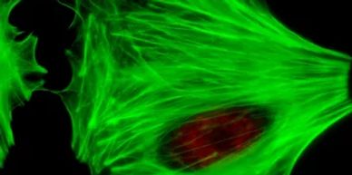
Similar to IHC, immunofluorescent stains highlight antigens with a fluorescent dye. We offer an array of IF stains to support and enhance your projects.
Plastic Processing (Hard Tissue/Devices)
Ground Section (EXAKT)
Multiple Special Stains
Ground Section (EXAKT)

Ground Section (EXAKT System) is used for studies with metal devices in the tissue that cannot be cut in paraffin.
Thin Section
Multiple Special Stains
Ground Section (EXAKT)
Thin Section is commonly done on specimens that contain metals that are soft enough to be cut by a tungsten blade. This yields a cleaner section for specimens that need to be embedded in plastic.
Multiple Special Stains
Multiple Special Stains
Multiple Special Stains
We use Spurr's resin which allows for full de-plasticization so we can perform nearly any special stain on plastic processed and embedded tissues.
Additional Services
Archiving
Pathology Consulting
Pathology Consulting
Long term storage of your study materials per GLP requirements (wet tissues, blocks, slides, paperwork, etc.)
Pathology Consulting
Pathology Consulting
Pathology Consulting
We offer pathologist oversight and report generation from board-certified veterinary pathologists.
Necropsy Support
Pathology Consulting
Necropsy Support
We have a team of trained necropsy personnel that can travel to your facility. We have experience from small to large animal models.
Slide Scanning
Device De-contamination
Necropsy Support
We offer digital imaging using our Leica Aperio CS2 or our Olympus VS200 whole slide scanners. Brightfield, polarizing light or immunofluorescent imaging is available for standard (1"x3" slides ) or large format (2"x3" slide).
Device De-contamination
Device De-contamination
Device De-contamination
Decontamination of devices after studies using glutaraldehyde as a cold sterilant.
Tissue Grossing
Device De-contamination
Device De-contamination
We have a trained team of technicians who can trim your specimens to the exact precision your study requires.